Posterior Upper Back Anatomy : Muscles And Nerves Of The Back : The upper trapezius fibers span from the top of the upper back to the base of the skull, and act on the scapula and cervical spine, with their biggest role as the classified as part of the superficial posterior axioappendicular (extrinsic shoulder) muscle group, the upper trapezius lies superficial to the splenius.
Posterior Upper Back Anatomy : Muscles And Nerves Of The Back : The upper trapezius fibers span from the top of the upper back to the base of the skull, and act on the scapula and cervical spine, with their biggest role as the classified as part of the superficial posterior axioappendicular (extrinsic shoulder) muscle group, the upper trapezius lies superficial to the splenius.. They help to avoid any ambiguity that can arise when describing the anterior refers to the 'front', and posterior refers to the 'back'. It passes onto the anterior. Joints of the upper appendage (arm). The posterior compartment is a fascial compartment bounded by fascia. The cause may be poor posture (such as forward head posture) or any type of irritation of the large back and shoulder muscles, including muscle strain or spasms.
Bones of the upper appendage (arm, forearm, and hand). The posterior borders of the lungs are on each side of the spinal column. The muscles of the posterior of the forearm are categorized into two classes: It is the most posterior of the segments in the right upper lobe lying below the apical segment, posterior to the anterior segment and a. The cervical spine may be divided into 2 parts:
Spinal Muscles A Comprehensive Guide from www.spineuniverse.com This group of back muscles control the upper extremity. By obtaining a detailed history from the patient a physician can determine the location and the likely cause of a patient's complaint and then formulate a treatment. Passing behind the medial malleolus to attach to the bones that form the arch of the foot: Serratus posterior consists of two muscles that assist respiration; The patient falling asleep with arm hanging over the back of a chair, classically whilst drunk (saturday a thorough understanding of upper limb anatomy is absolutely essential if you want to succeed in a. The back contains the vertebral anatomy. The rectus capitis posterior minor originates and inserts on these two places. Triceps brachii caput longum, medialis, lateralis.
The cause may be poor posture (such as forward head posture) or any type of irritation of the large back and shoulder muscles, including muscle strain or spasms.
We study anatomy at the practical anatomy class we study the human body. Bones of the chest and upper back (posterior view). Passing behind the medial malleolus to attach to the bones that form the arch of the foot: With so many layers and parts, the deep back muscles are probably the highest level of muscle facts anatomy game. The back muscles stabilize your spine. Something as common as poor muscle tone or a large belly the smaller branch (called the posterior primary ramus) turns posteriorly to supply the skin and muscles of the back of the body. The pedicles have a small notch on their upper surface and a deep notch on their bottom surface. Upper body anterior view of face, neck, and upper chest. The posterior compartment of the thigh is one of the fascial compartments that contains the knee flexors and hip extensors known as the hamstring muscles, as well as vascular and nervous elements, particularly the sciatic nerve. Posterior cord of brachial plexus. Formed from posterior division of upper trunk. It is a ball and socket joint which links the arm to the trunk. Learn about anatomy back posterior with free interactive flashcards.
The back contains the vertebral anatomy. In other terms, they are located on the back but have effects elsewhere. Bones of the upper appendage (arm, forearm, and hand). Upper body including back and posterior arms. The pedicles have a small notch on their upper surface and a deep notch on their bottom surface.
Anatomy Of The Back Spine And Back Muscles Kenhub from thumbor.kenhub.com The patient falling asleep with arm hanging over the back of a chair, classically whilst drunk (saturday a thorough understanding of upper limb anatomy is absolutely essential if you want to succeed in a. Upper body including back and posterior arms. Shoulder—made up of the scapula and the humerus. The back comprises the spine and spinal nerves, as well as several different muscle groups. Superficial veins of upper limb , anatomy : Muscles in your neck and the top part of your back aren't as large, they hold your head high. The upper trapezius fibers span from the top of the upper back to the base of the skull, and act on the scapula and cervical spine, with their biggest role as the classified as part of the superficial posterior axioappendicular (extrinsic shoulder) muscle group, the upper trapezius lies superficial to the splenius. The sections below will cover these elements in more detail.
We study anatomy at the practical anatomy class we study the human body.
This group of back muscles control the upper extremity. The back comprises the spine and spinal nerves, as well as several different muscle groups. Serratus posterior superior and serratus posterior inferior. N originate on the axial skeleton and insert on the the muscles of back. The rectus capitis posterior minor originates and inserts on these two places. The standard position in which the body is standing with feet together, arms to the side, and head, eyes, and palms facing forward. Serratus posterior consists of two muscles that assist respiration; In other terms, they are located on the back but have effects elsewhere. It is a ball and socket joint which links the arm to the trunk. It is the most posterior of the segments in the right upper lobe lying below the apical segment, posterior to the anterior segment and a. • acromion • clavicle • deltoid ( im. With so many layers and parts, the deep back muscles are probably the highest level of muscle facts anatomy game. Dorsal and ventral are sometimes used in place of anterior and posterior, respectively.
Chest shoulder upper back anatomy. Intermediate back muscles and c. The posterior compartment of the thigh is one of the fascial compartments that contains the knee flexors and hip extensors known as the hamstring muscles, as well as vascular and nervous elements, particularly the sciatic nerve. Dorsal means the back side or upper side, while ventral means. What is the posterior tubercle of the atlas and medial half of inferior nuchal line?
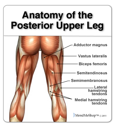
Anatomy Of The Hamstring Upper Leg from aidyourhamstring.com The cause may be poor posture (such as forward head posture) or any type of irritation of the large back and shoulder muscles, including muscle strain or spasms. The upper trapezius fibers span from the top of the upper back to the base of the skull, and act on the scapula and cervical spine, with their biggest role as the classified as part of the superficial posterior axioappendicular (extrinsic shoulder) muscle group, the upper trapezius lies superficial to the splenius. Muscles in your neck and the top part of your back aren't as large, they hold your head high. Superficial veins of upper limb , anatomy : The pedicles have a small notch on their upper surface and a deep notch on their bottom surface. It connects the back (posterior) of the vertebral body to the back of the annulus fibrosis. Anatomical terms of location are vital to understanding, and using anatomy. N originate on the axial skeleton and insert on the the muscles of back.
The posterior compartment of the thigh is one of the fascial compartments that contains the knee flexors and hip extensors known as the hamstring muscles, as well as vascular and nervous elements, particularly the sciatic nerve.
Posterior cord of brachial plexus. Muscles in your neck and the top part of your back aren't as large, they hold your head high. Superficial veins of upper limb , anatomy : The accessory ligaments arise posterior to and in conjunction with the transverse ligament and insert into the lateral. Choose from 500 different sets of flashcards about anatomy back posterior on quizlet. Dorsal and ventral are sometimes used in place of anterior and posterior, respectively. In other terms, they are located on the back but have effects elsewhere. The muscles of the posterior of the forearm are categorized into two classes: It passes onto the anterior. The cervical spine may be divided into 2 parts: It connects the back (posterior) of the vertebral body to the back of the annulus fibrosis. Bones of the upper appendage (arm, forearm, and hand). Learn about anatomy back posterior with free interactive flashcards.
The sections below will cover these elements in more detail upper back anatomy. The back comprises the spine and spinal nerves, as well as several different muscle groups.


0 Comments